

Applications
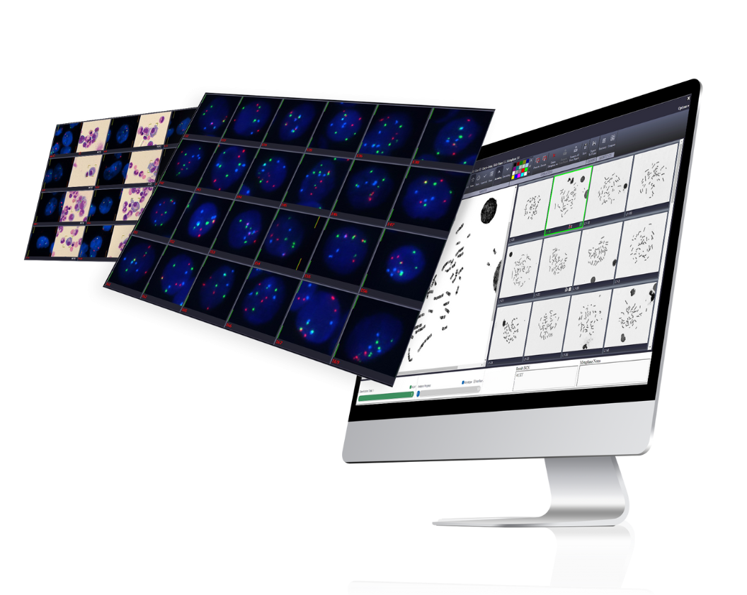
Automated Metaphases Imaging
Introducing streamlined, reliable, and technologically advanced imaging techniques for detection, segmentation, and user assisted classification of metaphases. Offering cutting-edge tools to simplify and expedite image review and on-screen analysis. With various solutions fitted to the laboratory sample volume, desired automation-level, and integrated web application for both local and remote use, BioView’s products are the ideal solution for your laboratory needs.
The information presented above does not constitute clinical utility. Please refer to the full intended use statement
FISH
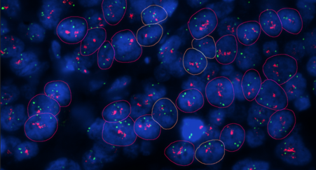
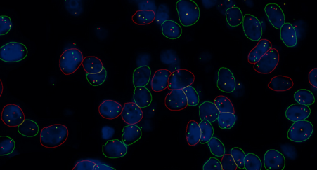
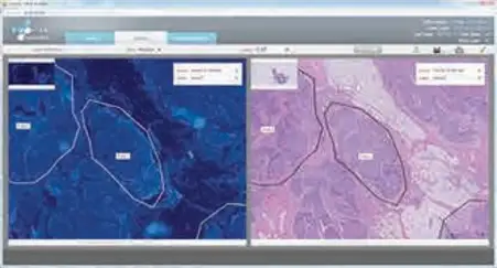
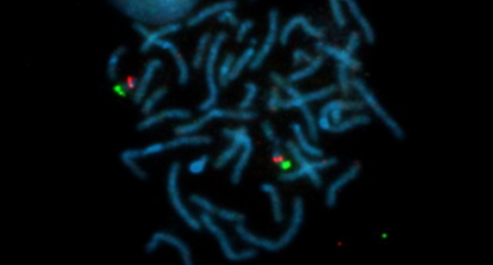
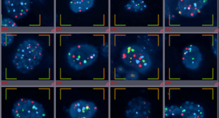
Rapid imaging and on-screen review of FFPE sections hybridized with FISH probes as well as matching between parallel tissue sections for targeted analysis.
Target - FISH
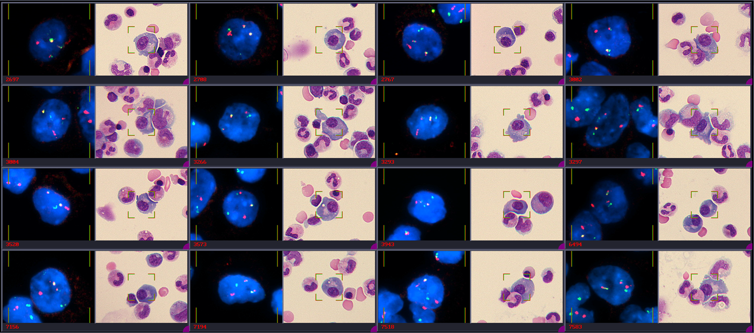
Automated imaging and matching of of multiple stains / markers of the same cell such as Giemsa + FISH, IHC + FISH, IFL +FISH. The platform performs unassisted relocation, automated FISH imaging and side-by-side presentation of the same cells processed using different stains.
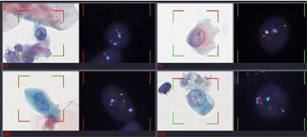
Rare cells detection
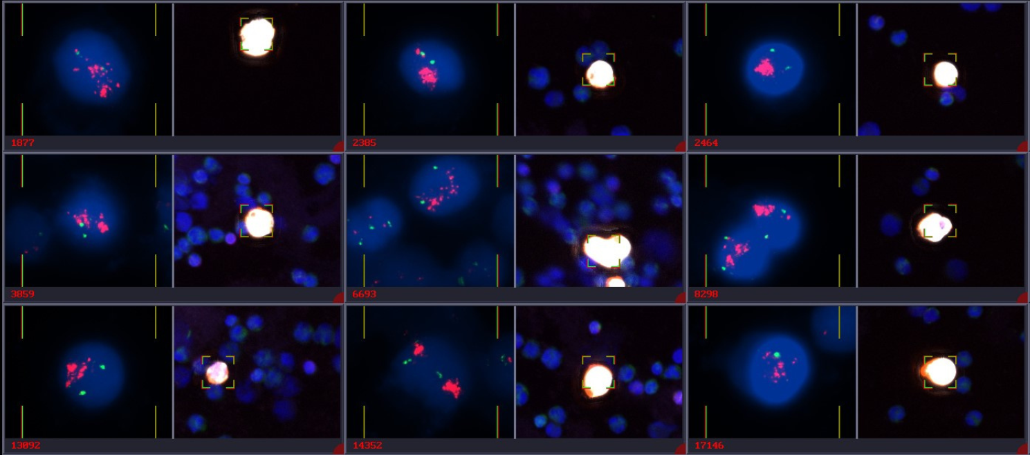
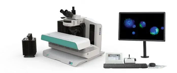
All-in-one automated platforms for rapid detection, imaging and characterization of rare cells from peripheral blood.
Powered by state-of-the-art imaging algorithms, the platform seamlessly integrates with any sample processing technique. Its sophisticated algorithms automatically detect and characterize rare cells regardless of the cell enrichment method or biomarker used, transforming what was once a manual process into a high-throughput automated workflow.
Custom-fitted to various microfluidic chip and cell filtration devices
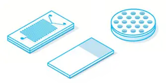
Designed to support automated imaging of various cell enrichment techniques independent of specific geometric patterns, 3D structures, and materials.
Fully adaptable to all fluorescent markers
Supports imaging & review of any combination of fluorescent markers, Proteins / RNA / DNA FISH, simultaneously or consecutively hybridized.
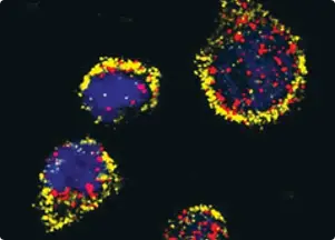
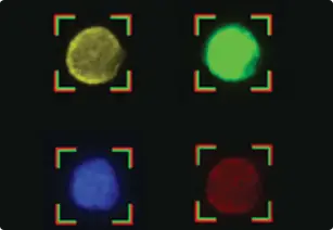
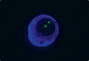
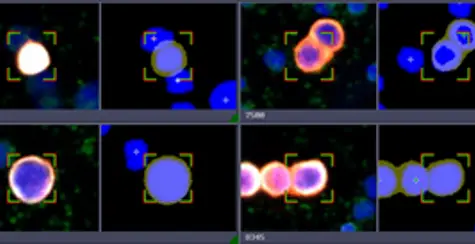
Automated Rare Cell detection
Cells are sorted and classified by combining advanced algorithms with fully adjustable gating criteria including morphology and signal attributes, ensuring precise detection of rare cell events within a sample while maintaining high specificity.
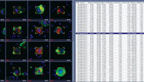
Cells characterization
The combined algorithms and biometric data allows forming assay-specific rare-cells gating criteria as well as assisting in assay development. Biometric data can be seamlessly exported to Excel spread sheet.
Multiplex imaging
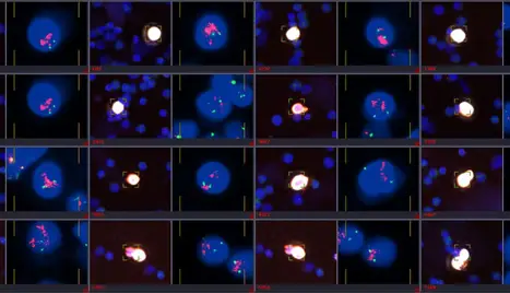
Utilizing the system automated cell relocation and capture, allows review of multiple biomarkers stained and imaged in consecutive sessions.
High throughput
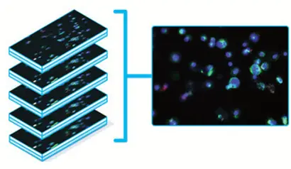
Combination of best in class optics and unique imaging algorithms specifically tailored to facilitate rapid 3D capture of all cells within the sample.
The information presented above does not constitute clinical utility. Please refer to the full intended use statement